Jerusalem, 11 November, 2026 (TPS-IL) — Israeli and US researchers have developed a blood-based technique that could reveal not only how much small-cell lung cancer a patient has, but also which genes cancer cells are actively using. The approach could help doctors better monitor disease changes and understand treatment response.
Unlike many existing liquid biopsies, which detect mutations in tumor DNA, the new method examines molecular signals showing which genetic instructions cancer cells use. The findings could pave the way for more precise treatments.
Lung cancer is the world’s most commonly diagnosed cancer and the leading cause of cancer-related deaths. Approximately 2.5 million people were diagnosed worldwide in 2022, accounting for 12.4% of new cancer cases, while 1.8 million died from the disease.
Small-cell lung cancer accounts for approximately 15% of lung cancer cases. It typically grows and spreads rapidly, and many patients are diagnosed at an advanced stage. Although tumors often initially shrink with treatment, the disease frequently returns. Obtaining new tumor tissue to track these changes can be difficult.
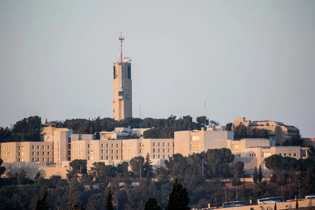
The international team was led by Prof. Nir Friedman and Dr. Gavriel Fialkoff of the Hebrew University-Hadassah Medical School, together with Dr. Nobuyuki Takahashi and Dr. Anish Thomas of the US National Cancer Institute, part of the National Institutes of Health (NIH). The findings were published in the peer-reviewed Journal of Clinical Investigation.
A New Way to Read Cancer’s Genetic Signals
The researchers examined microscopic fragments released into the bloodstream by dying cells. These include pieces of DNA wrapped around proteins called histones. Chemical marks on the histones indicate which genes were active in the cells that released the fragments.
Using cell-free chromatin immunoprecipitation sequencing (cfChIP-seq), the team analyzed these fragments to investigate cancer cells’ biological activity. Unlike conventional genetic testing, which detects alterations in DNA, the method reveals how cells use their genetic code.
The researchers analyzed 441 blood plasma samples from people with small-cell lung cancer, other cancers and healthy individuals. They compared blood-based signals with gene activity in tumor tissue. The procedure required only about one milliliter of plasma.
The team identified molecular patterns characteristic of small-cell lung cancer and developed a numerical score measuring their strength. The score correlated with tumor burden, as measured by medical imaging, and changed during treatment. Median scores declined following treatment, consistent with reduced tumor mass. Higher scores were also associated with shorter survival.
But the technique’s potential extends beyond measuring tumor size.
Small-cell lung cancer is not a uniform disease. Its tumors can be divided into molecular subtypes, defined by distinct patterns of gene activity. These differences may explain why patients respond differently to treatment and could become important as therapies targeting specific subtypes emerge.
The researchers found that blood-based molecular signals closely reflected the activity of key genes in patients’ tumor tissue. Some samples contained signals associated with multiple subtypes, suggesting that the technique could capture biological differences among cancer cells at different locations.
This could offer an advantage over conventional biopsies, which provide information from only one location. Blood can carry material released by tumors at multiple sites, potentially giving doctors a broader picture of the disease.
If validated in further studies, the test could help doctors monitor treatment response, track changes in tumor biology and identify cancer subtypes throughout the body, potentially reducing the need for repeated invasive biopsies. The researchers stressed that the test needs further study before doctors can use it to guide treatment or replace biopsies.